
CENTRAL RAY ANGULATION
The angle of the x-ray beam, the average angle of projection of the central ray, is essential for successful use of the bisecting exposure techniques. Both vertical and horizontal angulations must be considered.
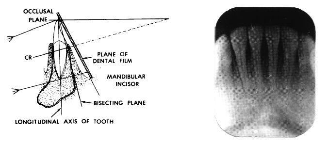
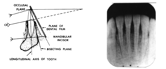
a. Vertical angulation is the up-and-down movement of the tube head or x-ray beam. The correct vertical angulation exists when the central ray is directed perpendicular to the bisector of the angle formed by the long axis of the tooth and the plane of the film (see figure 4-4). When this angulation is correct, the vertical dimension of the tooth will be as realistic as possible. Incorrect vertical angulation may cause two problems--foreshortening or elongation.
(1) Foreshortening exists if the vertical angulation is larger than necessary. The image of the teeth appears smaller than normal (see figure 4-5).
(2) Elongation exists if the vertical angulation is less than is necessary. The image of the teeth appears larger than normal (see figure 4-6).
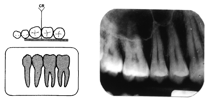
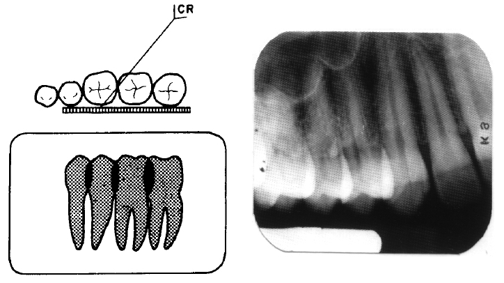
b. Horizontal angulation is the side-to-side movement of the tube head or x-ray beam. Correct horizontal angulation for successful radiographs exists when the central ray is perpendicular to the facial surfaces of the teeth and parallel to the mesial and distal surfaces (see figure 4-7). If the horizontal angulation is incorrect, overlapping will occur on the radiograph. Overlapping results when the proximal surfaces of adjacent


teeth are superimposed over one another (see figure 4-8). When this occurs, there will be a light area where the two teeth are overlapped or superimposed. The resultant light area is the inability of the x-ray beam to penetrate the two dense surfaces. Correct horizontal and vertical angulation of the x-ray beam is necessary to obtain radiographs of diagnostic quality.

